
Pain mechanism · Cranial nerve V
Trigeminal neuralgia
when a blood vessel becomes a pain generator
Classical trigeminal neuralgia is a clinical pain syndrome, not an imaging finding. Vascular contact becomes meaningful only when the phenotype, symptomatic side, compression site, and nerve deformation are concordant. Contact alone cannot establish causation or determine whether microvascular decompression is appropriate.
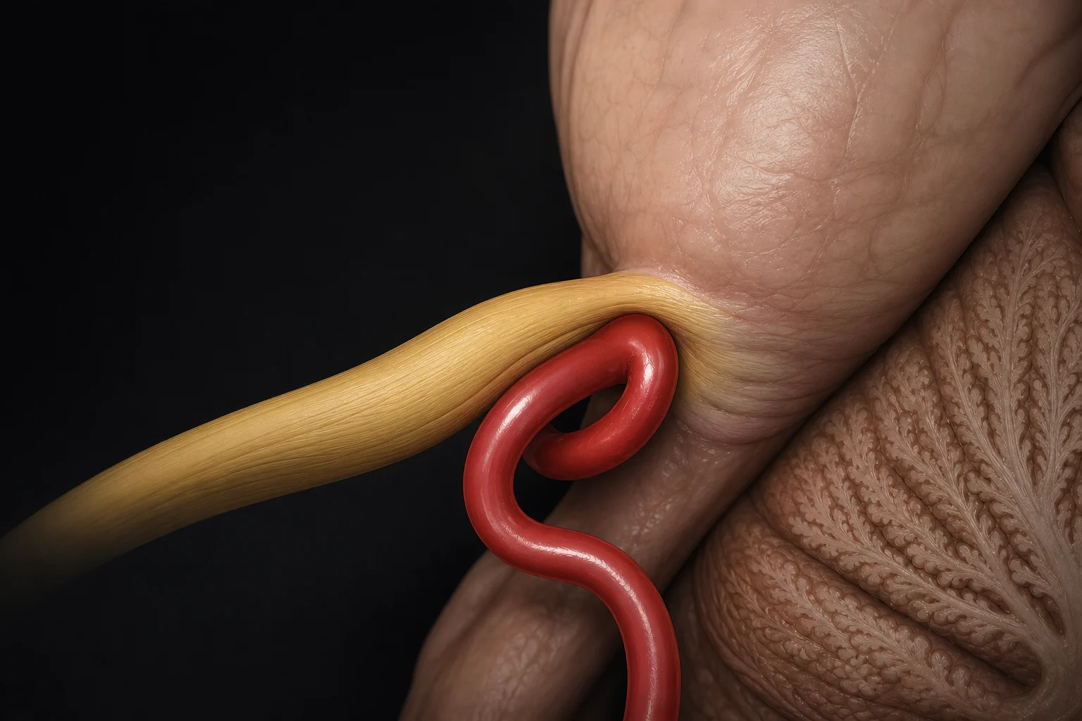 Pons
Trigeminal nerve · CN V
SCA loop
Focal compression near REZ
Pons
Trigeminal nerve · CN V
SCA loop
Focal compression near REZ
Core idea
The clinically important word is compression—not contact.
Neurovascular contact is common, including on asymptomatic nerves. Classical trigeminal neuralgia is more strongly supported when imaging or surgery shows compression at or near the root entry zone with morphological change such as displacement or atrophy of the nerve.
1. Mechanism: a focal conflict at the proximal trigeminal root
The trigeminal nerve carries facial sensation from the pons toward the trigeminal ganglion. In classical trigeminal neuralgia, an artery—most often the superior cerebellar artery in surgical and imaging series—may indent or displace the proximal cisternal nerve. AICA, veins, or more than one vessel can also be involved; the vessel must be identified from the individual anatomy rather than assumed from a diagram.
2. MRI: look for concordance and nerve deformation
MRI supports the diagnostic work-up and helps exclude secondary causes, but the diagnosis remains clinical. High-resolution heavily T2-weighted imaging, often assessed with angiographic sequences, is used to inspect the cisternal trigeminal nerves and adjacent vessels. The useful question is not simply “does a vessel touch the nerve?” but whether the relationship is on the symptomatic side, close to the root entry zone, and associated with indentation, displacement, or atrophy.
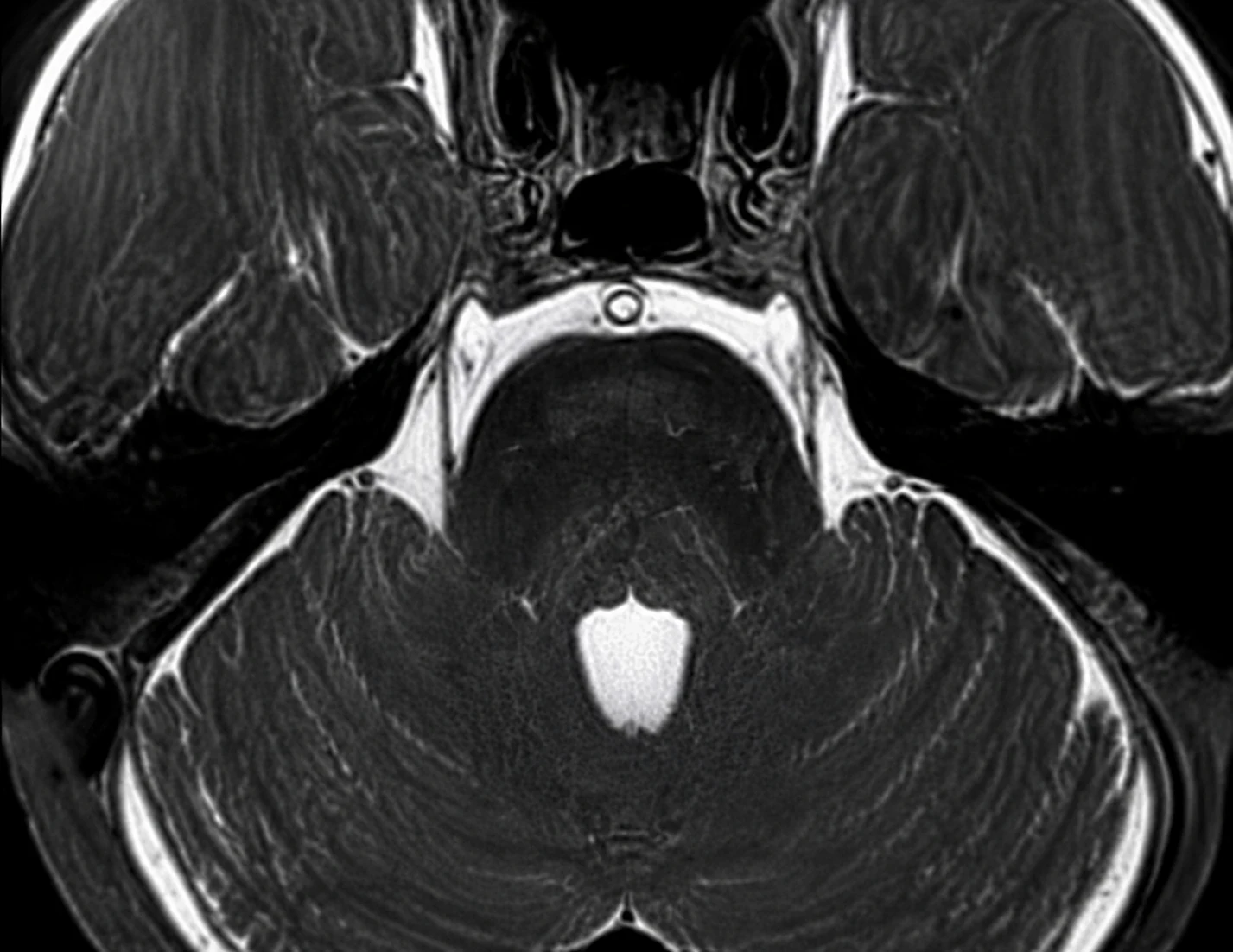 Pons
Inspect nerve shape, side, and REZ relationship
Pons
Inspect nerve shape, side, and REZ relationship
3. MVD: separate or reposition the responsible vessel
Microvascular decompression aims to relieve a concordant neurovascular conflict while preserving the trigeminal nerve. Depending on the operative anatomy, the vessel may be separated from the nerve with a small interposed material or moved and secured away from it. Figure 3 shows one simplified interposition concept; it is not a universal operative recipe.
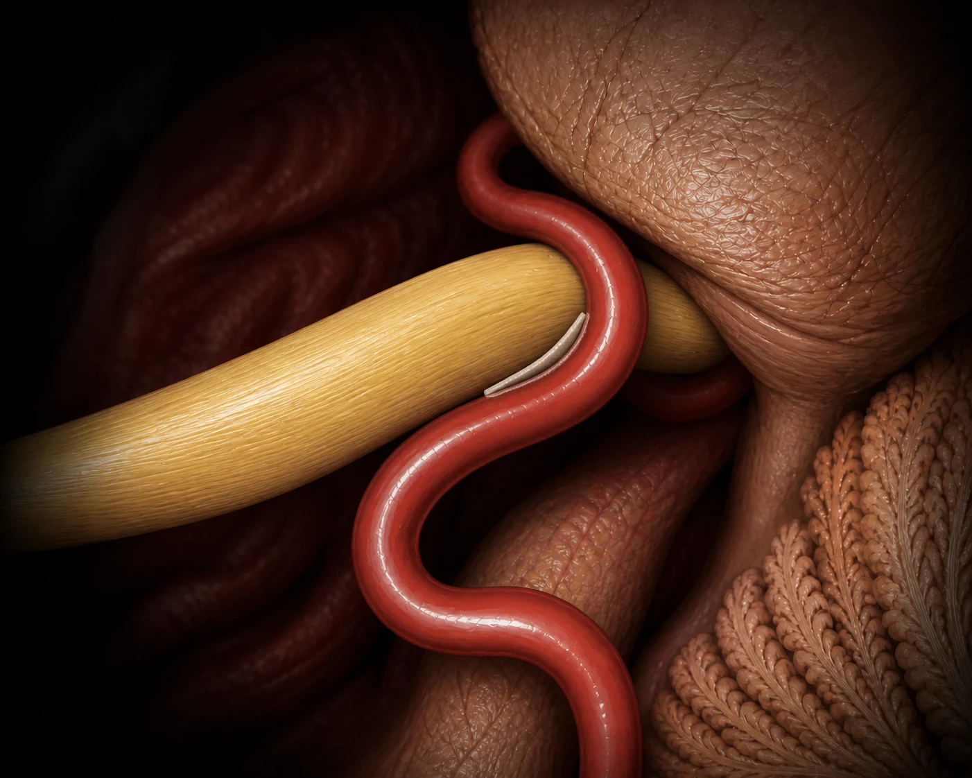 CN V
Repositioned vessel
One small interposition pledget
CN V
Repositioned vessel
One small interposition pledget
4. Decision boundary: an image is not an indication for surgery
Do not conclude
“A vessel touches the nerve, therefore it causes the pain.”
Contact can be incidental. Side, location, degree of compression, nerve morphology, and the pain phenotype must be assessed together.
Do not conclude
“A convincing MRI automatically means MVD.”
Treatment selection also depends on diagnostic certainty, medication response and tolerance, comorbidity, procedural risk, alternatives, and informed patient preference.
Use the image for
Understanding the clinical chain.
Symptoms define the syndrome; MRI tests the anatomical hypothesis; operative inspection confirms the relationship; decompression strategy follows the actual anatomy.
The strongest interpretation is concordant: typical pain, the same side, a plausible REZ conflict, and visible nerve deformation. Any one element in isolation is weaker.
Evidence summary
Clinical phenotype: classical trigeminal neuralgia is defined primarily by a characteristic unilateral, brief, triggerable, electric-shock-like pain syndrome. Imaging supports the anatomical hypothesis but does not replace the clinical diagnosis.
MRI interpretation: the superior cerebellar artery is a common culprit, but AICA, veins, or multiple vessels may also be involved. The most persuasive imaging pattern is a concordant conflict on the symptomatic side near the root entry zone with indentation, displacement, or atrophy. Contact without nerve deformation is common and cannot establish causation by itself.
Operative relevance: microvascular decompression separates or repositions the vessel while preserving trigeminal function. Interposition is one technique rather than a universal recipe; transposition or another strategy may be preferable depending on the operative anatomy, diagnostic certainty, procedural risk, alternatives, and informed patient preference.
References
- International Headache Society. ICHD-3: Classical trigeminal neuralgia.
- Bendtsen L, et al. European Academy of Neurology guideline on trigeminal neuralgia. Eur J Neurol. 2019.
- Cruccu G, et al. Trigeminal neuralgia: new classification and diagnostic grading for practice and research. Neurology. 2016.
- Maarbjerg S, et al. Significance of neurovascular contact in classical trigeminal neuralgia. Brain. 2015.
- Borges DJGN, et al. Evaluating the diagnostic role of MRI in cranial nerve hyperactive dysfunction syndromes: a controlled blinded study. Brain Spine. 2025.
- Akkara Y, et al. Incidence, role, and imaging-based diagnostic accuracy of neurovascular compression in trigeminal neuralgia. Br J Neurosurg. 2026.
- Outcome comparison between interposition and contactless transposition microvascular decompression approaches. J Neurosurg. 2025.
Review and provenance record
Editor reviewed — text and figures
Read the full review scope, dates, and limitations
- Editorial responsibility
- Neurosurgery.ai (site-name attribution; editor identity not published). Editorial identity.
- First published
- August 17, 2026
- Page updated
- September 6, 2026 — presentation or disclosure date; not an evidence or review date.
- Evidence search
- Not separately documented; no date inferred from publication or page maintenance.
- Editorial check
- The previous release records an English-language editorial update on August 30, 2026. No new literature appraisal is claimed for this disclosure update.
- Professional review
- The site editor reports completing professional review of the English article text on August 17, 2026, and confirmed the date and scope on September 6, 2026. This is editor self-review, not independent peer review.
- Reviewer attribution
- Neurosurgery.ai site editor (practicing neurosurgeon; identity not published)
- Confirmed review scope
- English article text and all three illustrations (Figures 1–3), including the AI-synthesized MRI-style image in Figure 2, as confirmed by the site editor. This is editor self-review, not independent peer review.
- Image-anatomy review
- The site editor confirms that the same review covered all three illustrations (Figures 1–3), including the synthetic MRI-style teaching image. This records the editor's anatomy review; no independent figure review or diagnostic validation is claimed.
- Professional-review date
- August 17, 2026
- Editor confirmation
- September 6, 2026 — confirms that review occurred; not the actual review date.
- Images
- Three AI-assisted images; Figure 2 is fully synthetic MRI, not a patient scan. The editor's scope confirmation covers the English text and all three figures currently presented. Existing v1 image files are unchanged and are separate from article version 1.3.
- Reviewer interests
- A reviewer-specific interests statement is not published. This must not be interpreted as a declaration of no conflicts.
- This update
- Version 1.3 records the editor-reported review date and text/figure scope, replacing the previous unspecified-attribution label. August 17 remains both the reported review date and first-publication date; September 6 is the confirmation/disclosure date. Medical narrative, scientific limitations and image files are unchanged; no new literature appraisal is claimed.