
Vascular mechanism · Spinal cord imaging
Spinal dural arteriovenous fistula
what 3D SPACE can show—and what it cannot prove
Heavily T2-weighted 3D SPACE can make serpiginous perimedullary vessels conspicuous and may help narrow a suspected shunt level. It remains a localization aid: catheter angiography is needed to define the arterial feeder, dural shunt and venous drainage for treatment planning.
Core idea
The visible vein is a clue—not necessarily the shunt.
An SDAVF is an arterial-to-venous connection at a dural nerve-root sleeve. The arterialized intradural draining vein can transmit pressure into a long segment of the perimedullary venous plexus. The most striking flow void, cord T2 hyperintensity or venous loop may therefore lie several vertebral levels away from the actual fistula.
1. The disease is venous-hypertensive myelopathy
In the usual thoracolumbar SDAVF, a radiculomeningeal arterial branch drains directly into a radicular vein within the dura at a spinal nerve-root sleeve. Arterial pressure is transmitted into the intradural venous system. The resulting venous hypertension reduces the pressure gradient available for normal cord drainage, producing venous congestive myelopathy and—if untreated—progressive neurological injury.
Routine MRI may show longitudinal cord T2 hyperintensity, cord swelling, enhancement and serpiginous perimedullary flow voids. None is independently diagnostic. The pattern should raise suspicion and prompt a vascular work-up rather than be treated as a direct map of the fistula.
2. Why heavily T2-weighted 3D SPACE can make abnormal veins visible
A heavily T2-weighted volumetric acquisition gives bright CSF a high-signal background. Rapid or complex blood flow may lose signal and appear as a dark tubular or serpiginous structure. Thin, near-isotropic source data can then be reviewed in multiple planes or with minimum-intensity projection, helping the reader follow a candidate draining vessel through the thecal sac.
This contrast is useful but conditional. Vessel conspicuity depends on flow velocity and direction, sequence parameters, motion, spatial resolution and reconstruction. A dark line is not automatically a vein, and an absent flow void does not exclude an SDAVF.
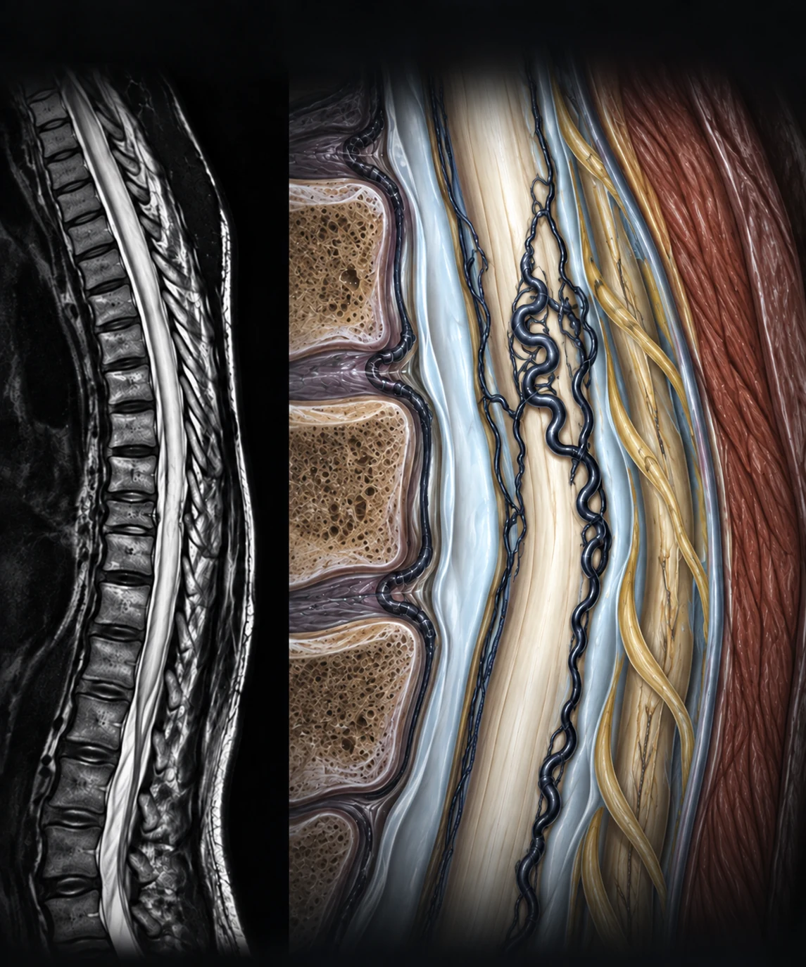
- 1Synthetic sagittal heavily T2-weighted 3D SPACE-style view
- 2Bright CSF background
- 3Spinal cord
- 4Dilated perimedullary veins rendered as low-signal flow voids
- 5Long-segment cord T2 hyperintensity compatible with venous congestion — not specific for reversible edema and does not localize the shunt
3. “Suggested shunt level” is the defensible wording
The original 3D SPACE study was a retrospective series of 16 patients with angiographically diagnosed SDAVF. Two blinded observers predicted the fistula site correctly in 81% and 88% of cases; accuracy reached 94% when a prediction within one vertebral level was accepted. This is a promising targeting result from a small selected cohort—not evidence that 3D SPACE can independently diagnose or exclude SDAVF.
Venous anatomy explains the limitation. In a separate 31-patient series, enlarged intrathecal draining veins extended a mean of 10 ± 7.7 spinal levels on angiography. A candidate vein approaching a root sleeve may narrow the search, but the longest, darkest or most congested segment does not necessarily mark the shunt.
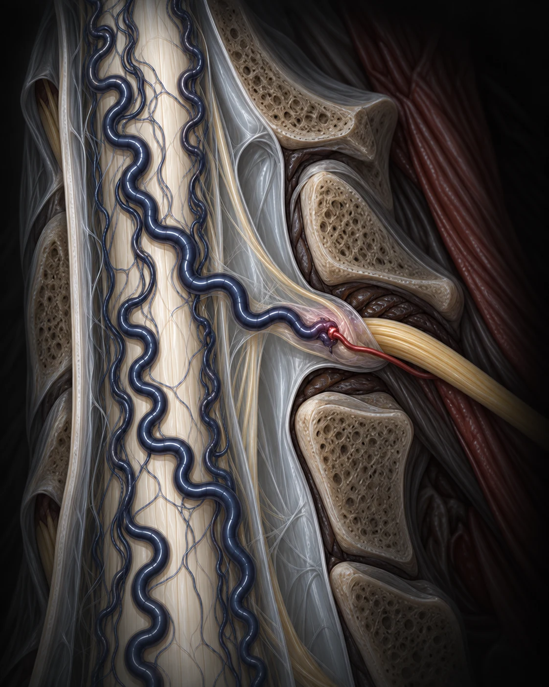
- 1Dilated perimedullary venous plexus
- 2Intradural radicular draining vein
- 3Arteriovenous shunt at the dural root sleeve
- 4Radiculomeningeal arterial feeder
4. MRA can guide the angiogram; it does not replace the angioarchitecture
Time-resolved or contrast-enhanced spinal MRA adds temporal and vascular information that noncontrast 3D SPACE does not provide. Recent reviews report high sensitivity and useful level prediction, but exact-level performance varies across small, heterogeneous studies. Those results cannot be transferred directly to a heavily T2-weighted structural sequence.
Complete spinal digital-subtraction angiography remains the reference examination for defining the arterial feeder, precise shunt, intradural venous drainage, adjacent critical arteries and a safe treatment strategy. It is not infallible: selected retrospective series document false-negative examinations when angiography was incomplete or technically limited. The practical model is integration—MRI raises suspicion, 3D SPACE or MRA may narrow the search, and angiography resolves the treatment anatomy.
Imaging workflow
Suspicion → localization → confirmation
Routine MRI may reveal a congestive myelopathy pattern. 3D SPACE and/or contrast-enhanced MRA may identify candidate abnormal vessels and a suspected level. Complete catheter angiography is then used to define the angioarchitecture and treatment target.
Evidence summary and selected sources
Evidence summary
| Evidence | What it supports | Boundary |
|---|---|---|
| 3D SPACE, 16 patients | Observer site prediction of 81% and 88%; 94% within one vertebral level. | Retrospective, selected, small; not a stand-alone diagnostic-accuracy study. |
| TR-MRA review, 4 studies | High pooled sensitivity; exact level in 59%, within one level in 97.4% of localized cases. | Wide uncertainty and protocol heterogeneity; not the same sequence as 3D SPACE. |
| 4D-MRA review | Correct side in 91% and level in 76%, supporting targeted angiography. | Does not establish that MRA defines every feeder or safely replaces DSA. |
| Venous-drainage series, 31 patients | Engorged intrathecal veins extended a mean of 10 ± 7.7 levels. | The most conspicuous vein may be remote from the fistula. |
| MRI/MRA series, 30 surgically confirmed cases | Routine MRI suggested SDAVF in all 30 cases; 15 underwent MRA, including cases investigated after negative DSA. | Retrospective, single-centre and surgically selected; it shows how incomplete studies can fail, not that MRA replaces complete DSA. |
| MRA versus DSA review, 13 studies and 133 patients | Pooled sensitivity favored MRA, while pooled localization accuracy favored DSA. | Small, heterogeneous, surgically confirmed cohorts; the pooled comparison does not validate noncontrast 3D SPACE. |
Do not conclude
“A flow void proves SDAVF.”
Flow voids are a vascular clue with mimics; the complete imaging and clinical context matters.
Do not conclude
“The largest vein marks the fistula.”
Arterialized venous drainage can extend many vertebral levels cranially or caudally.
Use the sequence for
Narrowing a difficult angiographic search.
When the vessel course is convincing, volumetric imaging may reduce—but not eliminate—the search space.
3D SPACE can help point to where the angiographer should look. It cannot declare that the search is over.
Selected references
- Kannath SK, et al. Utility of 3D SPACE T2-weighted volumetric sequence in the localization of spinal dural arteriovenous fistula. J Neurosurg Spine. 2016.
- Essibayi MA, et al. Spinal Dorsal Intradural Arteriovenous Fistulas: Natural History, Imaging, and Management. Neurology. 2023.
- Hetts SW, et al. Spinal dural arteriovenous fistulas and intrathecal venous drainage. J Neurosurg Spine. 2012.
- Wójtowicz K, et al. Usefulness of time-resolved MR angiography in spinal dural arteriovenous fistula. Neurosurg Rev. 2024.
- Raymaekers V, et al. Diagnostic Accuracy of 4D-MRA for the Detection and Localization of Spinal Dural Arteriovenous Fistulas. World Neurosurg. 2025.
- Saraf Lavi E, et al. The Role of MRA as a Preliminary Diagnostic Tool in Diagnosing and Localizing Spinal Dural Arteriovenous Fistula. AJNR Am J Neuroradiol. 2025.
- Khalafallah AM, et al. Evaluating the diagnostic accuracy of magnetic resonance angiography versus digital subtraction angiography for spinal dural arteriovenous fistulas. Neurosurg Rev. 2026.
Review and provenance record
Editor reviewed — text and figures
Read the full review scope, dates, and limitations
- Editorial responsibility
- Neurosurgery.ai (site-name attribution; editor identity not published). Editorial identity.
- First published
- September 15, 2026
- Page updated
- September 15, 2026 — presentation or disclosure date; not an evidence or review date.
- Evidence search
- September 15, 2026
- Editorial check
- Focused PubMed evidence search and source cross-check completed September 15, 2026. Eight unique source records informed the final editorial review; this is not a systematic review.
- Professional review
- The site editor completed professional review of the English article text and both figures on September 15, 2026. This is editor self-review, not independent peer review; separate neuroradiology or vascular-neurosurgery peer review is not claimed.
- Reviewer attribution
- Neurosurgery.ai site editor (practicing neurosurgeon; identity not published)
- Confirmed review scope
- Complete English article text and both AI-assisted synthetic illustrations (Figures 1–2), including the synthetic MRI-style panel in Figure 1, as confirmed by the site editor. This is editor self-review, not independent peer review.
- Image-anatomy review
- The editor review covered Figure 1 cord T2 signal, swelling and nonlocalizing presentation, and Figure 2 root sleeve, arterial feeder, fistulous point, intradural draining vein, nerve-root complex and perimedullary venous extent.
- Professional-review date
- September 15, 2026
- Editor confirmation
- September 15, 2026 — confirms that review occurred; not the actual review date.
- Images
- Two AI-assisted synthetic educational illustrations. Figure 1 includes a fully synthetic MRI-style panel, not a patient study. No patient data were used. The images are not validated for diagnosis, angiographic targeting or patient-specific treatment decisions.
- Reviewer interests
- A reviewer-specific interests statement is not published. This must not be interpreted as a declaration of no conflicts.
- This update
- Version 1.0 is the first public release. It distinguishes routine MRI, heavily T2-weighted 3D SPACE, contrast-enhanced MRA and complete DSA; describes 3D SPACE as a localization aid rather than definitive angiography; and records editor self-review of the complete text and both figures.