
Visual knowledge base
Neurosurgery Visual Atlas
A clinician-built visual knowledge base using AI-assisted medical illustration to explain how anatomy, mechanism, imaging, operative risk, and decision boundaries fit together. Clinician-built visual guides to anatomy, imaging, operative risk, and decision boundaries.
- Format
- AI-assisted illustration
- Review
- Status disclosed per page
- Boundary
- Educational, not patient-specific
AI-assisted illustration. Review status is shown per guide; no image is patient-specific or for diagnosis or surgical planning.
Atlas entries
Explore the visual guides.
Published entries carry a visible version, content-update date, and a review record distinguishing completed, confirmed, and pending work. Work in development remains clearly separated from reviewed or review-pending educational content.
Version and review status appear on every guide.
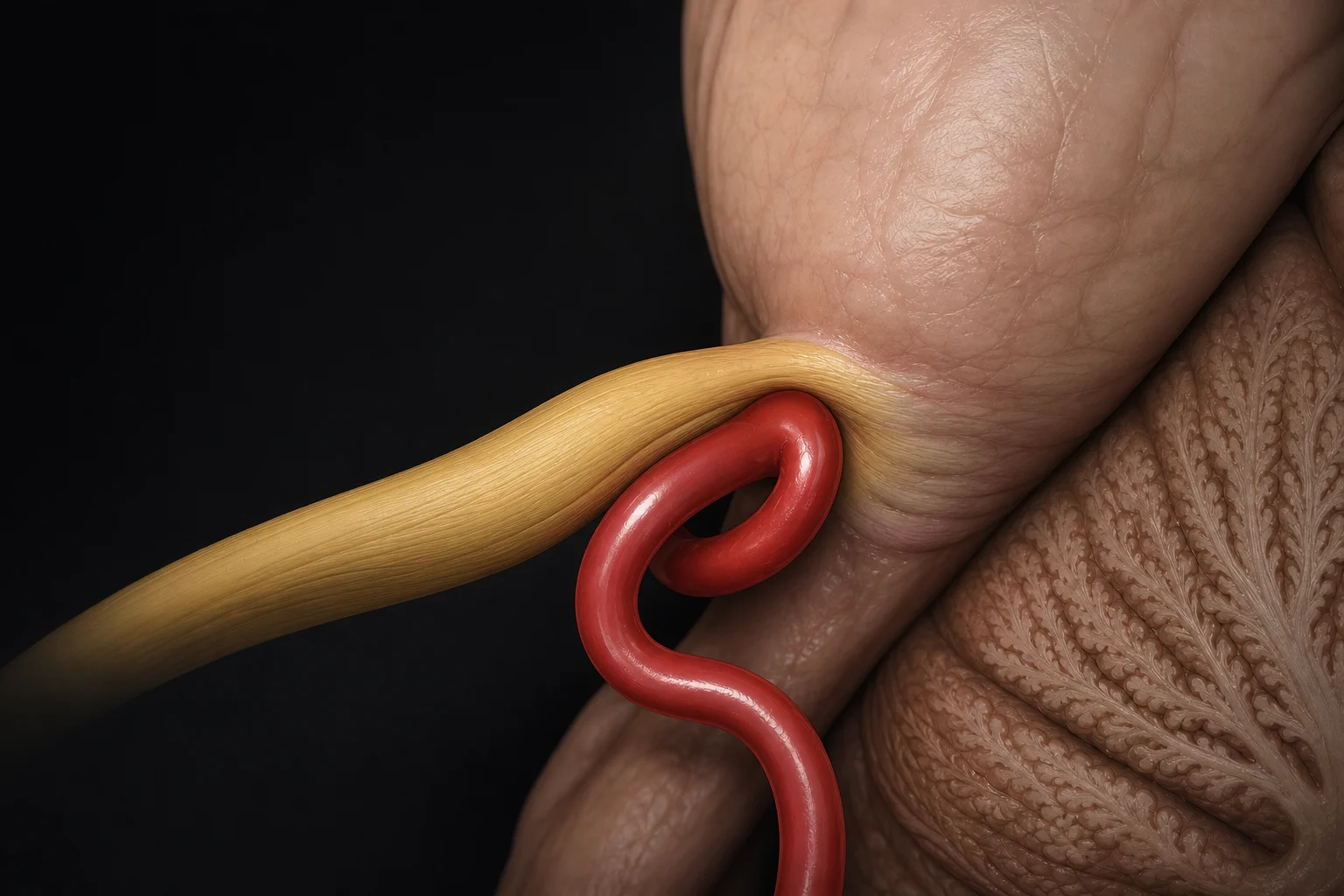 Illustration preview · Full-size figures inside
 AI-assisted illustration
Trigeminal neuralgia
Neurovascular compression, synthetic MRI teaching views, and the decision boundary for microvascular decompression.
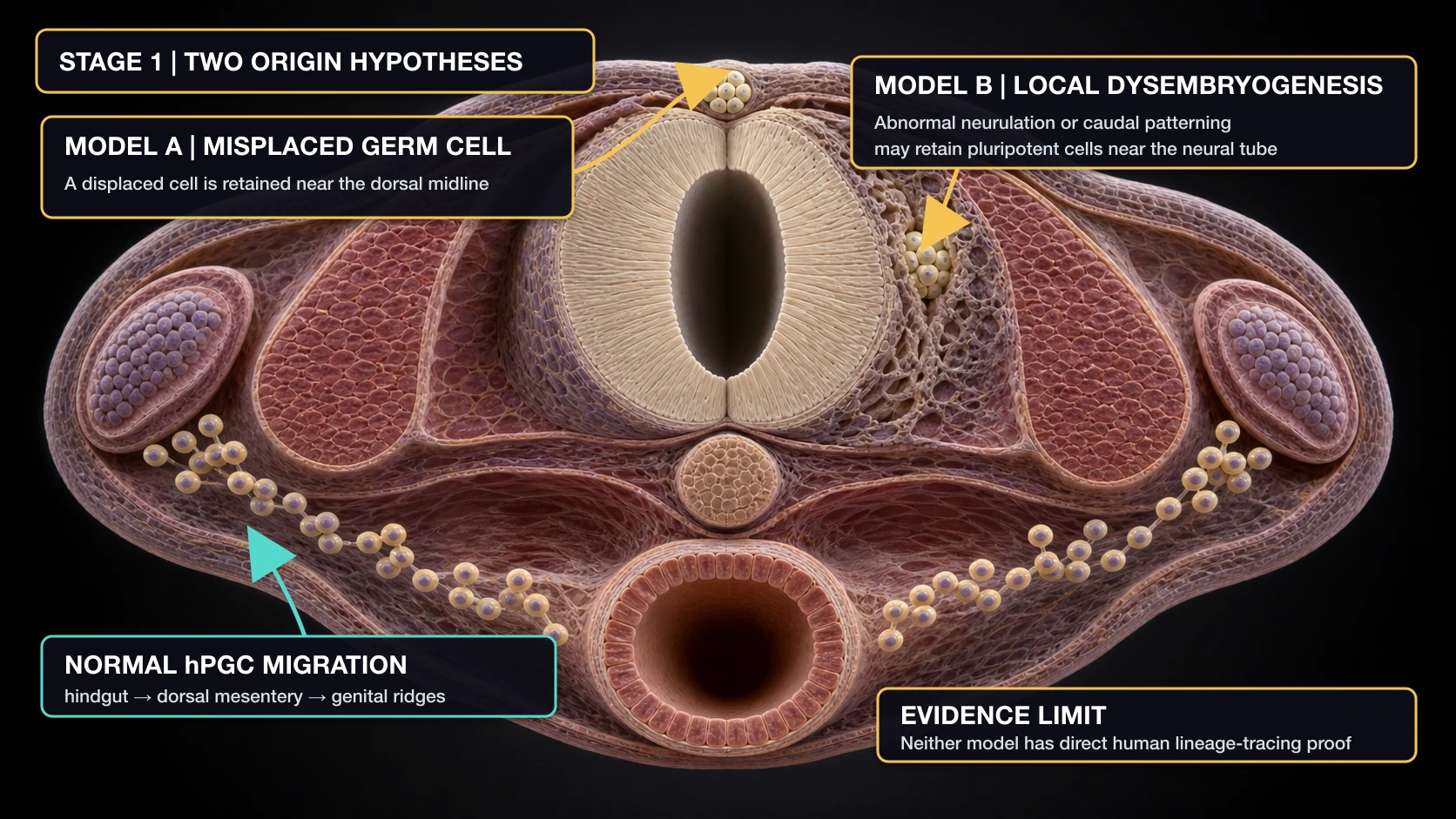 Illustration preview · Full-size figures inside
 AI-assisted illustration
Intramedullary spinal teratoma
Developmental hypotheses, cyst expansion, and the tumor–cord interface that can limit safe resection.
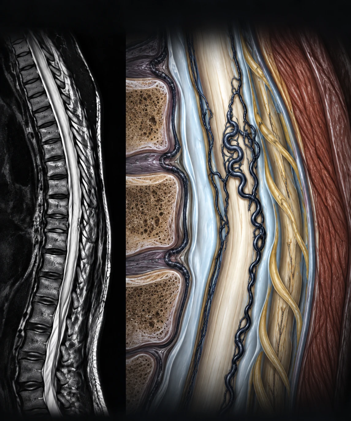 Illustration preview · Full-size figures inside
 AI-assisted illustration
Spinal dural arteriovenous fistula
Venous congestive myelopathy, perimedullary flow voids, and why 3D SPACE can suggest—but not prove—the shunt level.
In development: operative risk anatomy and monitoring. Unpublished work is not part of the reviewed Atlas.
Page architecture
What every Atlas page must answer.
The goal is not a gallery of attractive anatomy. Each sequence should clarify one clinical question from first mechanism to final boundary.
- 01Clinical question
Define the uncertainty in one sentence.
- 02Visual mechanism
Show the anatomy or pathology that drives it.
- 03Evidence
Connect the schematic to imaging, monitoring, or literature.
- 04Operative relevance
Explain what the visual changes in planning or risk.
- 05Decision boundary
State what cannot be inferred and when to defer.
Transparent by design
The image is only the first layer.
Every published page will carry an AI-use disclosure, clinical review status, figure-level captions, source references, version number, and separate content-update and review dates.
View the Atlas standard