
Developmental mechanism · Intramedullary tumor
Intramedullary spinal teratoma
from developmental cell rest to an unsafe interface
A mature spinal teratoma can be histologically benign yet densely adherent to functional cord. An absent cleavage plane may reflect congenital tissue integration, reactive gliosis or fibrosis, and traversing microvessels; it does not by itself establish malignant invasion.
Core idea
No cleavage plane does not automatically mean malignant invasion.
A mature teratoma may be inseparable from the spinal cord because of developmental attachment, chronic reactive change, or shared microvasculature. These mechanisms are not mutually exclusive, and none can be inferred from adherence alone. The operative target is maximal safe decompression—not anatomical completeness at the cost of neurological function.
1. Developmental origin: two complementary, unproven models
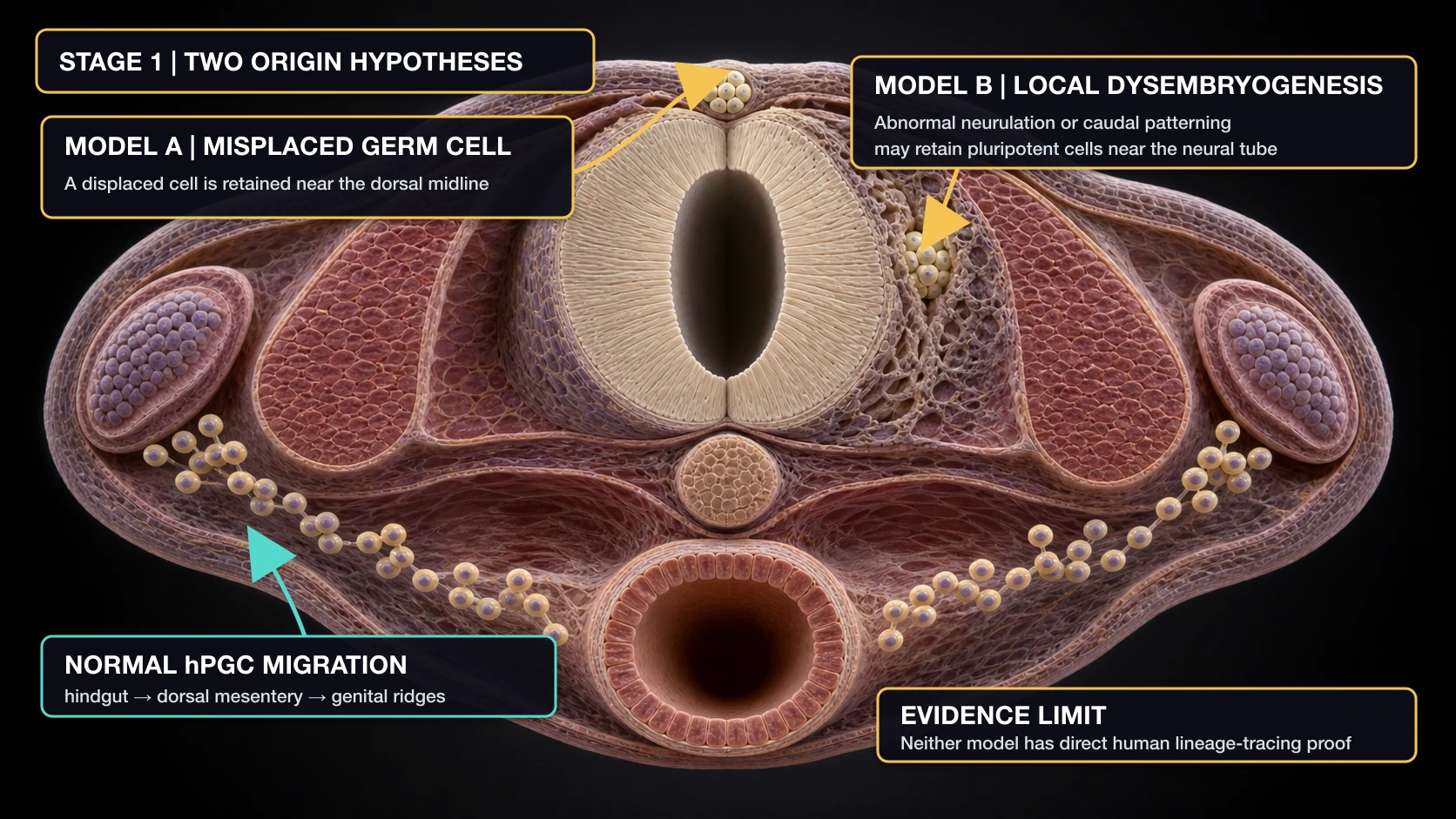
Human primordial germ cells are specified at approximately weeks 2–3, enter the hindgut region during early development, and from about week 5 traverse the dorsal mesentery toward the genital ridges. The misplaced germ-cell model proposes that an ectopic pluripotent germ cell is retained along or near a midline migratory route. The dysembryogenic model instead proposes that abnormal neurulation or caudal developmental-field signaling retains a local pluripotent cell population within or adjacent to the neural tube. Neither model has direct human lineage-tracing or molecular proof in spinal teratoma.
Midline location and cases without dysraphism are compatible with germ-cell misplacement, whereas the association with split cord malformation, tethering, spina bifida, myelomeningocele, dermal stigmata, and vertebral anomalies supports a broader developmental-field disturbance. Dysraphism is supportive, not required. A combined model—in which an abnormal embryonic environment retains either a misplaced germ cell or another pluripotent developmental cell—is a useful synthesis, but remains hypothetical.
2. A retained cell rest may develop with—not merely inside—the cord
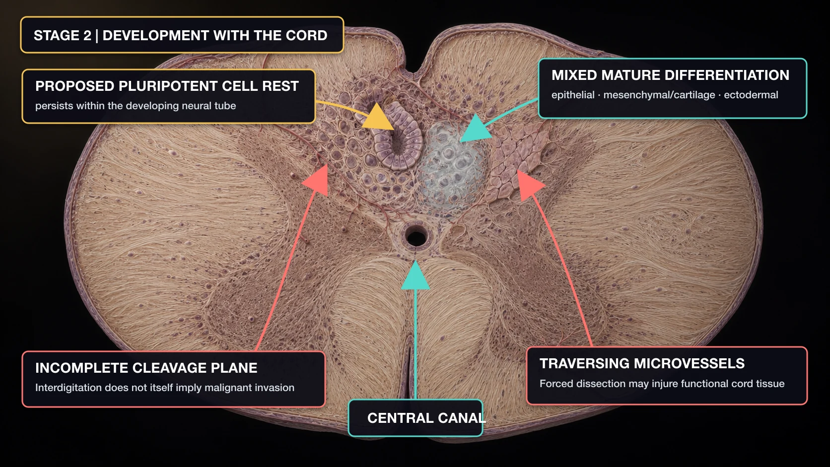
A proposed pluripotent cell rest can differentiate into epithelial, mesenchymal, neural, and skin-type tissues. If differentiation begins while the cord is organizing, congenital attachment could contribute to an indistinct margin or a capsule that is only partly separable. This developmental explanation is biologically plausible but has not been demonstrated by lineage studies.
Operative adherence can also be reinforced by capsular fibrosis, reactive gliosis, chronic inflammation, or prior microscopic hemorrhage. Interdigitation in a mature teratoma is therefore not synonymous with destructive malignant infiltration. Nevertheless, the surgical consequence can be similar: a safe plane may not exist, and vessels traversing the interface may supply both the lesion and functional cord.
3. Why a mature lesion can enlarge even when proliferation is slow
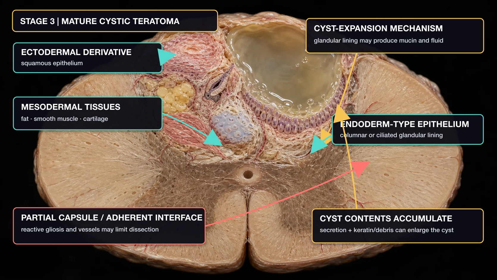
Mature teratomas contain well-differentiated derivatives of more than one embryonic germ layer. Squamous epithelium and neural or skin-type tissue represent ectodermal differentiation; fat, smooth muscle, cartilage, and vessels represent mesodermal differentiation; respiratory- or gastrointestinal-type glandular epithelium represents endodermal differentiation. The combination varies, and a limited specimen may not sample every component.
A cyst can enlarge through secretion and retention of material without rapid cellular proliferation. Glandular or columnar epithelium may produce mucin and fluid, while keratinizing squamous epithelium can add keratin and cellular debris. This mechanism is pathologically plausible and consistent with mucinous, fatty, or keratinous operative contents, but its quantitative contribution has not been measured in spinal teratoma cohorts.
4. Pathophysiology: slow expansion can still produce irreversible injury
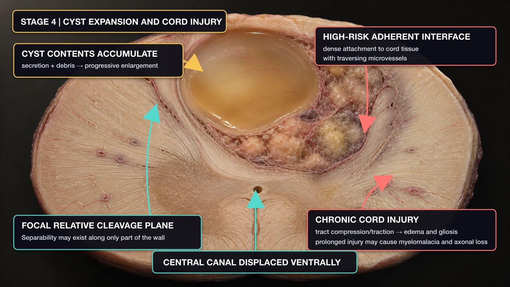
Progressive cyst enlargement and solid-tissue accumulation can expand the cord, displace and thin long tracts, distort local microcirculation, and move the central canal away from its usual position. Sustained compression or traction may produce edema, demyelination, gliosis, myelomalacia, and eventual axonal loss. Associated tethering or dysraphism can add a longitudinal traction component.
These mechanisms explain why symptoms may emerge only in adulthood despite a developmental origin: a small, slow-growing lesion can remain compensated for years until secretion, tissue accumulation, or tethering crosses a functional threshold.
5. Surgical boundary: maximal safe resection is not a euphemism
Do not conclude
“No plane means the lesion is malignant.”
A mature developmental lesion can be densely integrated with normal cord. Histopathology—not the visual impression of adherence—defines maturity and malignant components.
Do not conclude
“Electrocoagulation guarantees no recurrence.”
It may reduce secretion from treated lining, but residual solid or epithelial elements can remain viable, and durable recurrence data are sparse.
Use the image for
Understanding why a planned residual can be safer.
When functional cord and microvessels cross the interface, preserving neurological function can justify subtotal resection followed by long-term contrast-enhanced MRI surveillance.
Complete resection is preferred when a safe plane exists. When it does not, durable decompression with preserved function is a more meaningful endpoint than the word “total” in an operative note.
Evidence summary
Cell of origin: the misplaced primordial germ-cell and dysembryogenic models remain competing explanations. Embryology and associated dysraphic abnormalities make both plausible, but no spinal teratoma has been assigned to either lineage by direct human tracing.
Growth and cyst expansion: mature tissue can remain slow-growing while glandular secretion, keratin, and cellular debris accumulate. This explains how a histologically mature lesion may enlarge without implying aggressive proliferation. The relative contribution of each mechanism is not quantified in current series.
Extent of resection: observational evidence is inconsistent. A 146-case adult review associated complete resection with better outcomes and fewer recurrences, whereas a later review of 89 adult intramedullary mature teratomas found no statistically significant outcome difference by extent of resection and recorded only three recurrences. These case-based data are vulnerable to selection bias: more adherent lesions are both harder to remove and more likely to be left residual. The defensible principle is complete resection when safely achievable, otherwise maximal safe resection with long-term MRI follow-up.
References
- Kalani MYS, et al. Spinal intradural teratomas: developmental programs gone awry? Neurosurg Focus. 2012.
- Koen JL, McLendon RE, George TM. Intradural spinal teratoma: evidence for a dysembryogenic origin. J Neurosurg. 1998.
- Al-Sarraj S, et al. Clinicopathological study of seven cases of spinal cord teratoma: a possible germ cell origin. Histopathology. 1998.
- Wang L, et al. Intramedullary cervical spinal cord teratoma. Medicine (Baltimore). 2020.
- Chen Y, et al. Surgical treatment outcomes of patients with conus medullaris teratoma: a single-center experience of 39 patients. World Neurosurg. 2020.
- Prasad GL, Divya S. A comprehensive review of adult onset spinal teratomas: analysis of factors related to outcomes and recurrences. Eur Spine J. 2020.
- Paker B, et al. Adult-onset intramedullary teratomas: systematic review with outcome analysis. Acta Neurol Belg. 2025.
- Roelen BAJ, Chuva de Sousa Lopes SM. Stay on the road: from germ cell specification to gonadal colonization in mammals. Philos Trans R Soc Lond B Biol Sci. 2022.
- Ramakrishna NB, Alves-Lopes JP, Gruhn WH. Emerging principles and models of human primordial germ-cell development. Development. 2025.
Review and provenance record
Editor reviewed — text and figures
Read the full review scope, dates, and limitations
- Editorial responsibility
- Neurosurgery.ai (site-name attribution; editor identity not published). Editorial identity.
- First published
- August 30, 2026
- Page updated
- September 6, 2026 — presentation or disclosure date; not an evidence or review date.
- Evidence search
- Not separately documented; no date inferred from publication or page maintenance.
- Editorial check
- The previous release records literature-grounded editorial review on August 30, 2026. No new literature appraisal is claimed for this disclosure update.
- Professional review
- The site editor reports completing professional review of the English article text on August 28, 2026, and confirmed the date and scope on September 6, 2026. This is editor self-review, not independent peer review. Separate neuropathology and developmental-biology reviews are not documented.
- Reviewer attribution
- Neurosurgery.ai site editor (practicing neurosurgeon; identity not published)
- Confirmed review scope
- English article text and all four illustrations (Figures 1–4), as confirmed by the site editor. This is editor self-review, not independent peer review.
- Image-anatomy review
- The site editor confirms that the same review covered all four illustrations (Figures 1–4). This records the editor's anatomy review; no independent figure review is claimed.
- Professional-review date
- August 28, 2026
- Editor confirmation
- September 6, 2026 — confirms that review occurred; not the actual review date.
- Images
- Four AI-assisted schematics with English v2 annotation layers. The editor's scope confirmation covers the English text and all four figures currently presented. Image versions are separate from article version 1.3; the image files are unchanged.
- Reviewer interests
- A reviewer-specific interests statement is not published. This must not be interpreted as a declaration of no conflicts.
- This update
- Version 1.3 records the editor-reported review date and text/figure scope, replacing the previous pending label. August 28 is the reported review date; August 30 remains the first-publication date, and September 6 is the confirmation/disclosure date. Medical narrative, scientific limitations and image files are unchanged; no new literature appraisal is claimed.